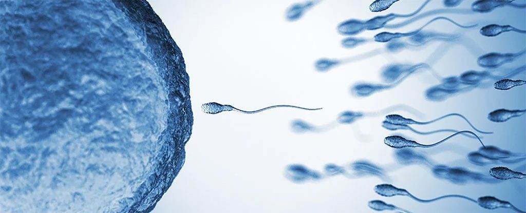
Según un estudio, los espermatozoides no se mueven como se creía hasta ahora
Una investigación publicada en Science Advances derribó la visión universalmente aceptada de cómo los espermatozoides “nadan”. Qué significa este descubrimiento
Más de trescientos años después de que Antonie van Leeuwenhoek usara uno de los primeros microscopios para describir el esperma humano como si tuviera una “cola que, al nadar, azota con un movimiento de serpiente, como anguilas en el agua”, los científicos han revelado que se trata de una ilusión óptica.
En este contexto, un nuevo estudio realizado por investigadores de la Universidad de Bristol en Reino Unido y la Universidad Nacional Autónoma de México, publicado en la revista Science Advances, ha dinamitado la visión universalmente aceptada de cómo los espermatozoides “nadan” al descubrir que su cola se mueve muy rápidamente en 3D, no de lado a lado en 2D como se pensaba.
Utilizando microscopía 3D y matemáticas de última generación, el doctor Hermes Gadelha, de la Universidad de Bristol, y los doctores Gabriel Corkidi y Alberto Darszon, de la Universidad Nacional Autónoma de México, han sido pioneros en la reconstrucción del verdadero movimiento de la cola del esperma en 3D.
Utilizando una cámara de alta velocidad capaz de grabar más de 55.000 cuadros en un segundo, y una platina de microscopio con un dispositivo piezoeléctrico para mover la muestra hacia arriba y hacia abajo a una velocidad increíblemente alta, pudieron escanear el esperma nadando libremente en 3D.
El innovador estudio revela que la cola del esperma es de hecho torpe y solo se mueve por un lado. Si bien esto debería significar que el golpe unilateral del espermatozoide lo haría nadar en círculos, los espermatozoides han encontrado una forma inteligente de adaptarse y nadar hacia adelante.
“Los espermatozoides humanos se daban cuenta si rodaban mientras nadaban, al igual que las nutrias juguetonas que sacaban sacacorchos a través del agua, su alimentación unilateral se normalizaría y nadarían hacia adelante”, explicó el doctor Gadelha, jefe del Laboratorio de Polímeros del Departamento de Matemáticas de Ingeniería de Bristol y experto en las matemáticas de la fertilidad.
De este modo, el estudio reveló que el giro rápido y altamente sincronizado de los espermatozoides causa una ilusión cuando se ve desde arriba con los microscopios 2D – la cola parece tener un movimiento simétrico de lado a lado, “como las anguilas en el agua”, como lo describió Leeuwenhoek en el siglo XVII.
“Sin embargo, nuestro descubrimiento muestra que los espermatozoides han desarrollado una técnica de natación para compensar su desequilibrio y al hacerlo han resuelto ingeniosamente un rompecabezas matemático a escala microscópica: creando simetría a partir de la asimetría”, apuntó Gadelha.
“El giro de los espermatozoides sin embargo complejo: la cabeza del espermatozoide gira al mismo tiempo que la cola del espermatozoide gira en la dirección de la natación. Esto se conoce en la física como precesión, muy parecido a cuando las órbitas de la Tierra y Marte giran alrededor del Sol”, enfatizó el investigador.
Los sistemas de análisis de semen asistidos por ordenador que se utilizan hoy en día, tanto en clínicas como en investigación, todavía usan visión en 2D para ver el movimiento de los espermatozoides. Por lo tanto, como el primer microscopio de Leeuwenhoek, todavía son propensos a esta ilusión de simetría mientras evalúan la calidad del semen. Este descubrimiento, con su novedoso uso de la tecnología del microscopio 3D combinado con las matemáticas, puede proporcionar una nueva esperanza para desbloquear los secretos de la reproducción humana.
“Dado que más de la mitad de la infertilidad se debe a factores masculinos, comprender la cola del espermatozoide humano es fundamental para desarrollar futuras herramientas de diagnóstico para identificar espermatozoides no saludables”, aseguró Gadelha, cuyo trabajo ha revelado anteriormente la biomecánica de la curvatura del espermatozoide y las precisas tendencias rítmicas que caracterizan el avance de un espermatozoide.
Los doctores Corkidi y Darszon fueron pioneros en la microscopía 3D para la natación de espermatozoides. “Esto fue una increíble sorpresa, y creemos que nuestro microscopio 3D de última generación revelará muchos más secretos ocultos en la naturaleza. Un día esta tecnología estará disponible para los centros clínicos”, dijo Corkidi.
“Este descubrimiento revolucionará nuestra comprensión de la motilidad del esperma y su impacto en la fertilización natural. Se sabe tan poco sobre el intrincado entorno del tracto reproductivo femenino y sobre cómo los espermatozoides que nadan influyen en la fertilización. Estas nuevas herramientas nos abren los ojos a las increíbles capacidades que tiene el esperma”, concluyó Darszon.
Con información Europa Press

Sperm don’t swim anything like we thought they did, new study finds
New research upends more than three centuries of beliefs about how sperm move.
Under a microscope, human sperm seem to swim like wiggling eels, tails gyrating to and fro as they seek an egg to fertilize.
But now, new 3D microscopy and high-speed video reveal that sperm don’t swim in this simple, symmetrical motion at all. Instead, they move with a rollicking spin that compensates for the fact that their tails actually beat only to one side.
“It’s almost like if you’re a swimmer, but you could only wiggle your leg to one side,” said study author Hermes Gadêlha, a mathematician at the University of Bristol in the U.K. “If you did this in a swimming pool and you only did this to one side, you would always swim in circles. … Nature in its wisdom came [up] with a very complex, ingenious way to go forward.”
Strange swimmers
The first person to observe human sperm close up was Antonie van Leeuwenhoek, a Dutch scientist known as the father of microbiology. In 1677, van Leeuwenhoek turned his newly developed microscope toward his own semen, seeing for the first time that the fluid was filled with tiny, wiggling cells.
Under a 2D microscope, it was clear that the sperm were propelled by tails, which seemed to wiggle side-to-side as the sperm head rotated. For the next 343 years, this was the understanding of how human sperm moved.
“[M]any scientists have postulated that there is likely to be a very important 3D element to how the sperm tail moves, but to date we have not had the technology to reliably make such measurements,” said Allan Pacey, a professor of andrology at the University of Sheffield in England, who was not involved in the research.
The new research is thus a “significant step forward,” Pacey wrote in an email to Live Science.
Gadêlha and his colleagues at the Universidad Nacional Autónoma de México started the research out of “blue-sky exploration,” Gadêlha said. Using microscopy techniques that allow for imaging in three dimensions and a high-speed camera that can capture 55,000 frames per second, they recorded human sperm swimming on a microscope slide.
“What we found was something utterly surprising, because it completely broke with our belief system,” Gadêlha told Live Science.
The sperm tails weren’t wiggling, whip-like, side-to-side. Instead, they could only beat in one direction. In order to wring forward motion out of this asymmetrical tail movement, the sperm head rotated with a jittery motion at the same time that the tail rotated.
The head rotation and the tail are actually two separate movements controlled by two different cellular mechanisms, Gadêlha said. But when they combine, the result is something like a spinning otter or a rotating drill bit. Over the course of a 360-degree rotation, the one-side tail movement evens out, adding up to forward propulsion.
“The sperm is not even swimming, the sperm is drilling into the fluid,” Gadêlha said.
The researchers published their findings today (July 31) in the journal Science Advances.
Asymmetry and fertility
In technical terms, how the sperm moves is called precession, meaning it rotates around an axis, but that axis of rotation is changing. The planets do this in their rotational journeys around the sun, but a more familiar example might be a spinning top, which wobbles and dances about the floor as it rotates on its tip.
“It’s important to note that on their journey to the egg that sperm will swim through a much more complex environment than the drop of fluid in which they were observed for this study,” Pacey said. “In the woman’s body, they will have to swim in narrow channels of very sticky fluid in the cervix, walls of undulating cells in the fallopian tubes, as well have to cope with muscular contractions and fluid being pushed along (by the wafting tops of cells called cilia) in the opposite direction to where they want to go. However, if they are indeed able to drill their way forward, I can now see in much better clarity how sperm might cope with this assault course in order to reach the egg and be able to get inside it,” Pacey said
Sperm motility, or ability to move, is one of the key metrics fertility doctors look at when assessing male fertility, Gadêlha said. The rolling of the sperm’s head isn’t currently considered in any of these metrics, but it’s possible that further study could reveal certain defects that disrupt this rotation, and thus stymy the sperm’s movement.
Fertility clinics use 2D microscopy, and more work is needed to find out if 3D microscopy could benefit their analysis, Pacey said.
“Certainly, any 3D approach would have to be quick, cheap and automated to have any clinical value,” he said. “But regardless of this, this paper is certainly a step in the right direction.”
Originally published in Live Science.


Debe estar conectado para enviar un comentario.